
Muscular atrophy, outlined because the lack of muscle tissue, is a critical subject for immobilized sufferers on Earth and for people throughout spaceflight, the place microgravity prevents regular muscle loading. In vitro modeling is a vital step in understanding atrophy mechanisms and testing countermeasures earlier than animal trials.
The most perfect atmosphere for modeling should be empirically decided to finest mimic recognized responses in vivo. To simulate microgravity situations, murine C2C12 myoblasts have been cultured in a rotary cell tradition system (RCCS). Alginate encapsulation was in contrast towards polystyrene microcarrier beads as a substrate for culturing these adherent muscle cells.
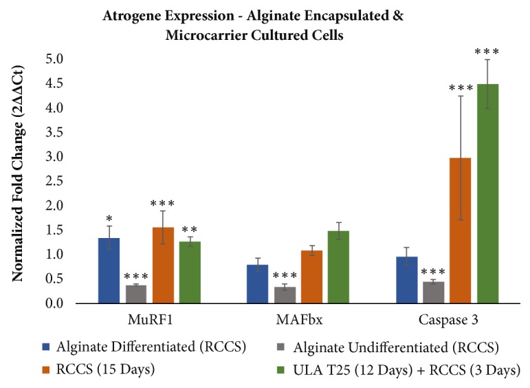
Changes after tradition underneath simulated microgravity have been characterised by assessing mRNA expression of MuRF1, MAFbx, Caspase 3, Akt2, mTOR, Ankrd1, and Foxo3. Protein focus of myosin heavy chain 4 (Myh4) was used as a differentiation marker. Cell morphology and substrate construction have been evaluated with brightfield and fluorescent imaging.
Differentiated C2C12 cells encapsulated in alginate had a big enhance in MuRF1 solely following simulated microgravity tradition and have been morphologically dissimilar to regular cultured muscle tissue. On the opposite hand, C2C12 cells cultured on polystyrene microcarriers had considerably elevated expression of MuRF1, Caspase 3, and Foxo3 and simply identifiable multinucleated myotubes.
The extent of differentiation was larger in simulated microgravity and protein synthesis extra lively with elevated Myh4, Akt2, and mTOR. The in vitro microcarrier mannequin described herein considerably will increase expression of a number of of the identical atrophy markers as in vivo fashions.
However, in contrast to animal fashions, MAFbx and Ankrd1 weren’t considerably elevated and the fold change in MuRF1 and Foxo3 was decrease than anticipated. Using a normal commercially obtainable RCCS, the substrates and tradition strategies described solely partially mannequin adjustments in mRNAs related to atrophy in vivo.

Assessment of NAD+metabolism in human cell cultures, erythrocytes, cerebrospinal fluid and primate skeletal muscle.
The reduction-oxidation state of NAD+/NADH is vital for mobile well being with NAD+ and its metabolites taking part in vital roles in getting older and pathologies. Given the inherent autooxidation of decreased dinucleotides (i.e. NADH/NADPH), and the well-established differential stability, the correct measurement of NAD+ and its metabolites is technically difficult.
Moreover, pattern processing, normalization and measurement methods can profoundly alter outcomes. Here we developed a fast and delicate liquid chromatography mass spectrometry-based methodology to quantify the NAD+ metabolome with cautious consideration of those intrinsic chemical instabilities.
Utilizing this methodology we assess NAD+ metabolite stabilities and decide the presence and concentrations of NAD+ metabolites in clinically related human samples together with cerebrospinal fluid, erythrocytes, and primate skeletal muscle.