A multicenter comparison of quantification methods for antisense oligonucleotide-induced DMD exon 51 skipping in Duchenne muscular dystrophy cell cultures.
Duchenne muscular dystrophy is a deadly illness brought on by lack of dystrophin. Skipping of exons adjoining to out-of-frame deletions has confirmed to revive dystrophin expression in Duchenne sufferers.
Exon 51 has been essentially the most studied goal in each preclinical and medical settings and the provision of standardized procedures to quantify exon skipping could be advantageous for the analysis of preclinical and medical knowledge.
OBJECTIVE
To evaluate methods at present used to quantify antisense oligonucleotide-induced exon 51 skipping in the DMD transcript and to offer steering in regards to the methodology to make use of.
METHODS
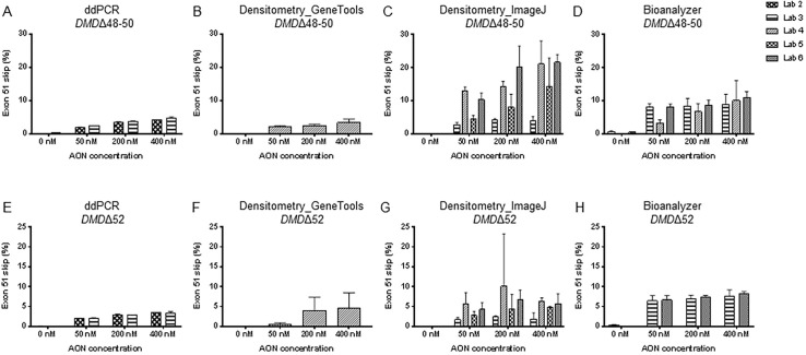
Six laboratories shared blinded RNA samples from Duchenne patient-derived muscle cells handled with completely different quantities of exon 51 concentrating on antisense oligonucleotide. Exon 51 skipping ranges have been quantified utilizing 5 completely different strategies: digital droplet PCR, single PCR assessed with Agilent bioanalyzer, nested PCR with agarose gel picture evaluation by both ImageJ or GeneTools software program and quantitative real-time PCR.
RESULTS
Differences in imply exon skipping ranges and dispersion across the imply have been noticed throughout the completely different strategies. Results obtained by digital droplet PCR have been reproducible and confirmed the smallest dispersion. Exon skipping quantification with the opposite methods confirmed overestimation of exon skipping or excessive knowledge variation.
CONCLUSIONS
Our outcomes counsel that digital droplet PCR was essentially the most exact and quantitative methodology. The quantification of exon 51 skipping by Agilent bioanalyzer after a single spherical of PCR was the second-best selection with a 2.3-fold overestimation of exon 51 skipping ranges in comparison with digital droplet PCR.
A multicenter comparison of quantification methods for antisense oligonucleotide-induced DMD exon 51 skipping in Duchenne muscular dystrophy cell cultures.
A mechanism for semaphorin-induced apoptosis: DNA injury of endothelial and myogenic cells in major cultures from skeletal muscle.
One hallmark of most cancers is its capability to recruit a vascular provide to assist speedy development. Suppression of angiogenesis holds potential as a second-line or adjuvant remedy to stunt most cancers development, development, metastasis, and post-resection regeneration.
To start to check the speculation that semaphorin 3A and 3F collectively, will induce endothelial cell apoptosis by inducing DNA injury, combined major cultures remoted from regular grownup mouse skeletal muscle have been handled for 48 hr with Sema3A ± Sema3F (100ng/mL).
Changes in surviving-cell density, DNA synthesis, DNA restore (gamma-Histone 2AX, γH2AX, an oblique measure for DNA injury), and apoptotic DNA fragmentation (TUNEL staining) have been assayed in cultures of CD31+ endothelial and desmin+ muscle cells. Sema3F elevated DNA damage-associated DNA restore in each cell varieties. Co-treatment with Sema3A+3F elevated γH2AX staining ~25-fold over management ranges, and additional elevated apoptosis in comparison with management and Sema3A alone.
Results have been negated by therapy with neutralizing anti-semaphorin antibodies and are interpreted as suggesting that Sema3A might sensitize endothelial however not muscle cells to Sema3F-induced DNA injury. These preliminary findings on a posh system of interacting cells might contribute to growing purposes that would goal angiogenic regulatory mechanisms for their therapeutic potential towards most cancers development and metastasis.